Ripping up the nuclear envelope Biology Diagrams An in vitro nuclear disassembly system reveals a role for the RanGTPase system and microtubule-dependent steps in nuclear envelope breakdown. J. Cell Biol. 178 , 595-610 (2007).

The COPI complex functions in nuclear envelope breakdown and is recruited by the nucleoporin Nup153. Dev Cell. 2003;5:487-498. doi: 10.1016/s1534-5807(03)00262-4. This paper implicates the Golgi COPI vesiculation machinery in breakdown of the nuclear envelope. In addition, COPI recruitment to the nuclear envelope is facilitated by its

Orchestrating nuclear envelope disassembly and reassembly during ... Biology Diagrams
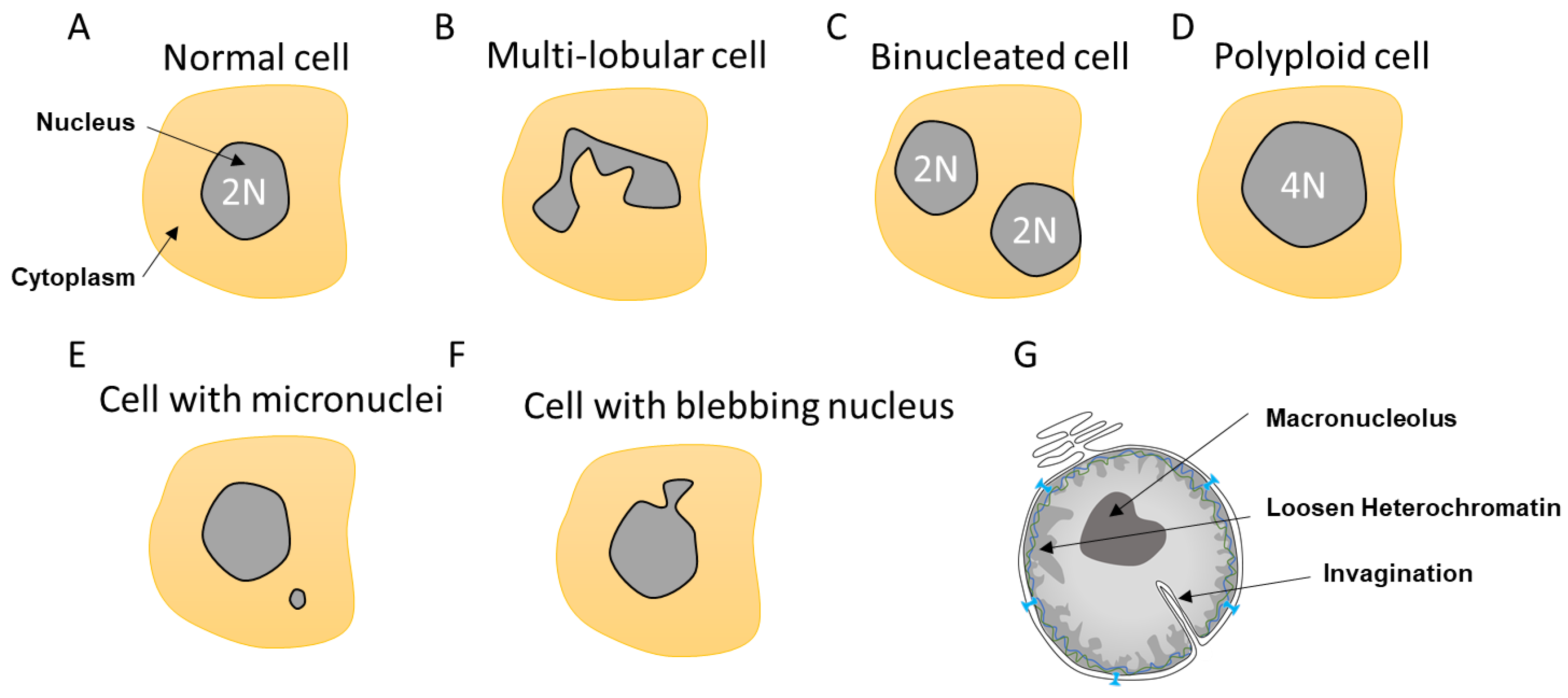
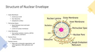
The nuclear envelope is punctured by around a thousand nuclear pore complexes, about 100 nm across, with an inner channel about 40 nm wide. [9] Aberrant nuclear envelope breakdown has also been observed in laminopathies and in cancer cells leading to mislocalization of cellular proteins, the formation of micronuclei and genomic instability.

Through this process, the nuclear envelope projects tubules that capture damaged DNA, mediating its repair. Current models suggest that DNA double-strand breaks (DSBs) can move to the nuclear
Mechanisms and functions of nuclear envelope remodelling Biology Diagrams
The mechanism of nuclear envelope breakdown (NEBD) was investigated in live cells. Early spindle microtubules caused folds and invaginations in the NE up to one hour prior to NEBD, creating mechanical tension in the nuclear lamina. The first gap in the NE appeared before lamin B depolymerization, at the site of maximal tension, by a tearing mechanism. Nuclear envelope breakdown was investigated during meiotic maturation of starfish oocytes. Fluorescent 70-kDa dextran entry, as monitored by confocal microscopy, consists of two phases, a slow uniform increase and then a massive wave. From quantitative analysis of the first phase of dextran entry, and from imaging of green fluorescent protein chimeras, we conclude that nuclear pore disassembly